
Figure 1.
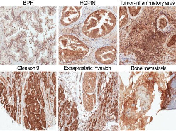
Immunohistochemical staining of GRM1 in normal and malignant prostate tissues. BPH shows nuclear staining in basal cells and absence of staining in normal luminal acinar cells. Moderate to intense cytoplasmic staining is noted in HGPIN. Intense nuclear and cytoplasmic staining is noted in tissue infiltrating macrophages, stromal cells and PCa cells within the tumor-inflammatory area. Intense cytoplasmic staining was observed in malignant luminal acinar cells of a Gleason 9 (5+4) tumor, and in tumor cells invading into extraprostatic adipose and muscle tissues. Intense anti-GRM1 staining was noted in scattered metastatic PCa cells within fragmented pieces of bone. Original magnification: ×200. BPH, benign prostate hyperplasia; GRM1, metabotropic glutamate receptor 1; HGPIN, high-grade prostate-intraepithelial neoplasia; PCa, prostate cancer.