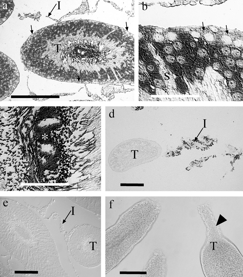
Figure 12.

Immunocytochemistries for endogenous glutamate and for 𝒹-aspartate. Glutamate immunoreactivity (a–c) was associated with somata in the outer two-thirds of the tubules (T), though the most superficial cells (arrows), presumed to be spermatogonia, tended to be weakly labeled. Radially oriented Sertoli cells (S) were frequently unlabeled or very weakly labeled. Strong labeling of the lumen of the tubules suggested that high levels of glutamate were present in the sperm and in amorphous deposits (*), which we interpret as representing glutamate that was fixed in the luminal fluid. Very little glutamate labeling was present in interstitial cells (I). 𝒹-aspartate uptake (d) was evident in interstitial cells (I) but not in the tubules (T). Conversely, in control sections (e) not exposed to 𝒹-aspartate, the interstitial cells were unlabeled. Panel f demonstrates that the cut ends of the isolated tubules were typically sealed (arrowhead) due to constriction of the tubules, thereby preventing ingress of 𝒹-aspartate. Scale bars: a, d, e, f=100 µm; b, c=25 µm.
