
Figure 2.
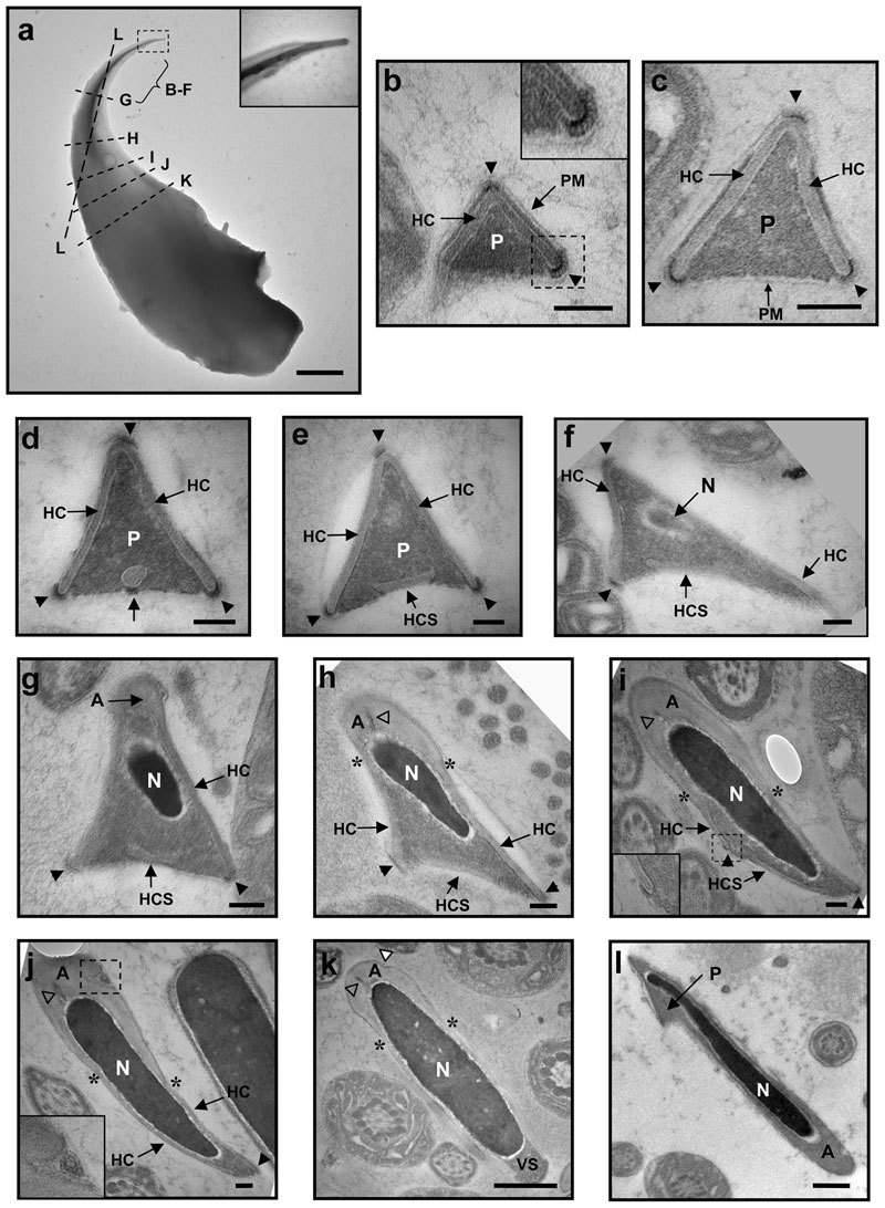
Electron micrographs of mouse cauda epididymidal sperm. (a) Image of a whole-mount sperm head. The bracket marks the region from where cross-sections in b–f are derived, whereas the broken lines correspond to the planes of cross-sections in g–l. The cross-sections are localized on the sperm head based on their longitudinal lengths. In b–k, the dorsal edges of the perforatorial triangles point to the top, whereas in l, the apical tip of the hook points to the top-left corner. Arrowheads in b–j point to the HRs at the apexes; asterisks in h–k locate the limits between the acrosome cap and the head cap; open triangles in h–k point to the indents inside the acrosome, and the white triangle in k points to the indent on the acrosome surface. In a, b, i and j, the boxed regions are enlarged in the insets. For a, scale bar=1 µm; for b–j, scale bar=100 nm; for k and l, scale bar=500 nm. A, acrosome cap; HC, head cap; HCS, separated head cap segment; N, nucleus; P, perforatorium; PM, plasma membrane; VS, ventral spur.