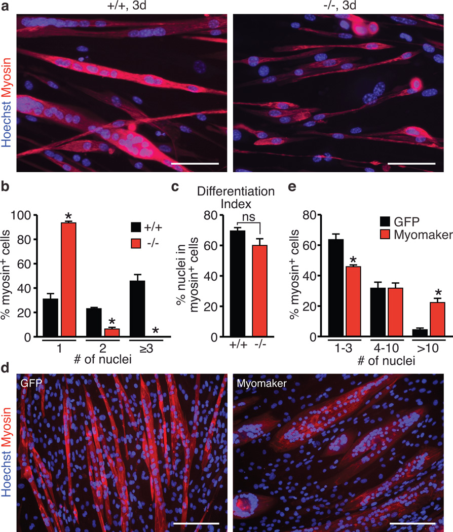
Figure 3. Control of myoblast fusion by Myomaker.
a, Myoblasts from WT (+/+) and Myomaker−/− E17 embryos were differentiated for 3 days, and stained for myosin and a nuclear stain (Hoechst). Myomaker−/− myoblasts failed to fuse. b, Quantitation of the number of nuclei present in a myosin+ cell indicates Myomaker−/− myoblasts cannot form myotubes with three or more nuclei. c, Differentiation index, calculated as the percentage of nuclei in myosin+ cells, indicated null myoblasts can activate the myogenic program. d, C2C12 cells infected with a retrovirus encoding GFP or Myomaker were induced to differentiate for 4 days then stained with a myosin antibody and Hoechst (nuclei). e, Quantitation of the percentage of myosin+ cells that contained the indicated number of nuclei. Quantification was performed after 3 days of differentiation in (b), (c), and after 4 days in (e). Scale bars: a, 100 µm e, 200 µm. Data are presented as mean ± SEM from three independent experiments. * P < 0.05 compared to +/+ in b, c or GFP-infected cells in e. ns in c is not statistically significant.

