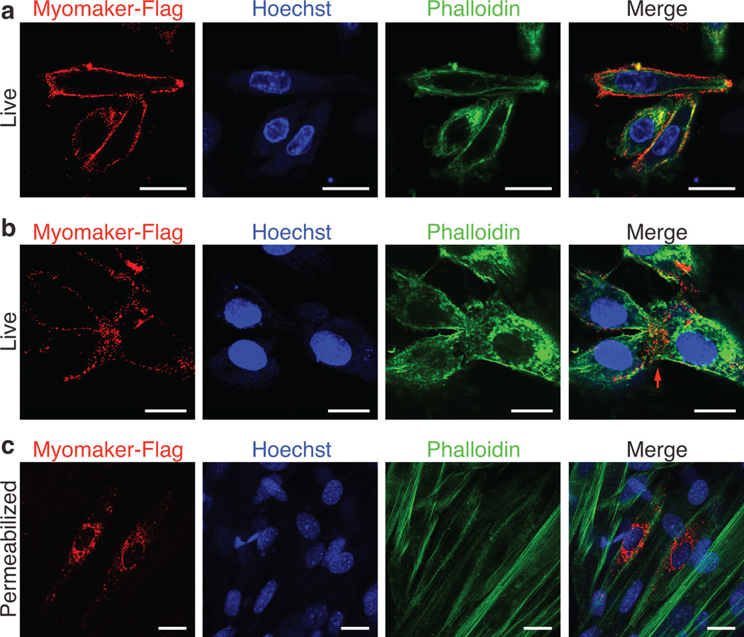
Figure 4. Myomaker is expressed on the cell membrane of myoblasts.
a, C2C12 cells were infected with Myomaker-Flag and live cells were stained 2 days after differentiation with Flag antibody on ice. After Flag staining, cells were then fixed and permeabilized and stained with Phalloidin (F-actin) and Hoechst (nuclei) to illustrate cell membrane localization of Myomaker-Flag. b, Cells were stained as in (a) to visualize Myomaker-Flag in fusing cultures. The red arrow depicts sites of cell-cell interaction. f, Myomaker-Flag infected C2C12 cells were fixed, permeabilized, and stained with Flag antibody, Phalloidin, and Hoechst showing the vesicle localization of the intracellular protein. Scale bars: 20 µm.

