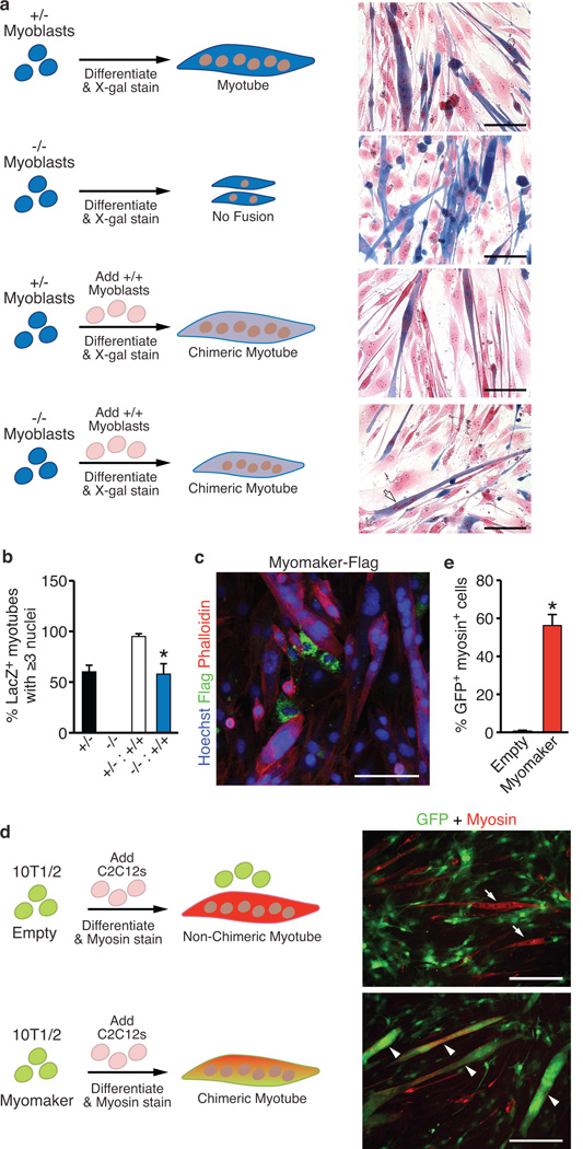
Figure 5. Myomaker participates in the myoblast membrane fusion reaction.
a, Myomaker+/− and Myomaker−/− myoblasts express LacZ, and were either plated alone or mixed with WT myoblasts, induced to differentiate for 4 days, and stained with X-gal and nuclear fast red to determine the amount of fusion. Myomaker+/− myoblasts, alone or in the presence of WT myoblasts fused normally, illustrated by myotubes with robust LacZ staining. Myomaker−/− myoblasts alone exhibited an inability to fuse. Addition of WT myoblasts to Myomaker−/− myoblasts resulted in chimeric myotubes (arrow) indicating fusion between the two cell populations. b, Quantitation of the percentage of LacZ+ myotubes containing ≥3 nuclei shows null myoblasts can only form myotubes with three or more nuclei in the presence of WT myoblasts. c, Phalloidin and Flag staining of C2C12 myoblasts after infection with Myomaker-Flag illustrates the lack of Flag staining in myotubes. d, 10T1/2 fibroblasts were infected with GFP-retrovirus and either Empty- or Myomaker-retrovirus and then mixed with C2C12 cells and differentiated. Myotube formation was monitored by myosin staining, and fusion of fibroblasts was determined by visualization of GFP in myosin+ myotubes. Myosin+ GFP+ myotubes (arrowheads) are evident in cultures containing Myomaker-infected fibroblasts, whereas myosin+ GFP− myotubes (arrows) were observed in Empty-infected cultures. e, Quantitation of the percentage of GFP+ fibroblasts, infected with Empty- or Myomaker-retrovirus, that fused to myosin+ myoblasts. Scale bars: a, 100 µm; c, 20 µm; d, 200 µm. Data are presented as mean ± SEM from three independent experiments. * P < 0.05 compared to −/− in b and compared to empty in e.

