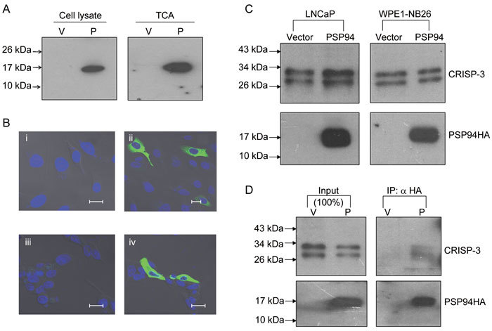
Figure 2.

Secreted recombinant PSP94 interacts with endogenous CRISP-3. (A): Western blot analysis for detection of PSP94HA. Cell lysate and TCA-precipitated proteins derived from the conditioned medium from PC3 cells that were transiently transfected with either empty vector (V) or a PSP94 expression construct (P) were probed with anti-HA antibody. (B): Localization of PSP94HA by immunofluorescence. PC3 cells that were transiently transfected with empty vector (i) and a PSP94 expression construct (ii) as well as LNCaP cells transiently transfected with empty vector (iii) and a PSP94 expression construct (iv) were first probed with polyclonal anti-HA antibody and then with FITC-labelled secondary antibody. Bars = 20 μm. (C): Effect of ectopically expressed PSP94 on endogenous CRISP-3. TCA-precipitated proteins from vector-transfected and PSP94 expression construct-transfected WPE1-NB26 and LNCaP cells were subjected to Western blot analysis 48 h after transfection. The upper panel was probed with anti-CRISP-3 antibody and the lower panel was probed with anti-HA antibody. (D): Co-immunoprecipitation of PSP94 and CRISP-3 from the conditioned medium of vector-transfected and PSP94 expression construct-transfected WPE1-NB26 cells. PSP94HA was immunoprecipitated using anti-HA agarose-conjugated beads. The upper portion of the Western blot was probed with anti-CRISP-3 antibody and the lower portion was probed with anti-HA antibody. The lanes marked with input show the total amount of CRISP-3 and PSP94HA that was immunoprecipitated from the conditioned medium.
