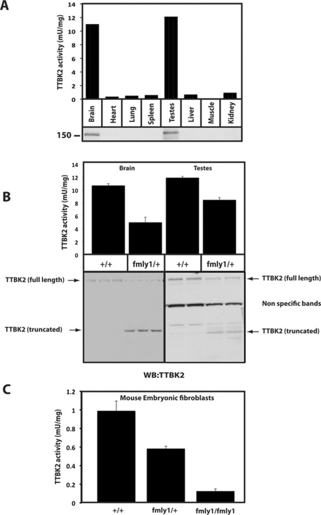
Figure 7. Study of TTBK2 in wild-type and TTBK2fmly1/+ knockin mice.
(A) The indicated tissue extracts were generated from wild-type mice. Extracts were immunoblotted for TTBK2 (lower panel) or TTBK2 was immunoprecipitated and subjected to a TTBK2 kinase assay employing the TTBKtide peptide substrate (upper panel). Results are means of duplicate experiments that were repeated four separate times with similar results. (B) Brain and testes lysates were generated from TTBK2+/+ and TTBK2fmly1/+ mice and subjected to immunoblot or TTBK2 kinase assay analysis, as in (A). (C) MEFs were generated from TTBK2+/+, TTBK2fmly1/+ and TTBK2fmly1/fmly1 E10 embryos as described in the Materials and methods section. TTBK2 activity was assessed following immunoprecipitation as in (A). Owing to the low levels of TTBK2 protein expressed in MEFs and high antibody background in immunoprecipitates, we were unable to detect expression of TTBK2 by immunoblot analysis. Results in (B) and (C) are means±S.D.

