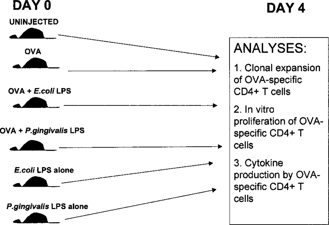
FIGURE 1.
Experimental design. B6.PL.Thy-1a (B6.PL) mice, or C57BL/6 mice that were reconstituted with OT-2 cells were injected with either soluble OVA, soluble OVA + E. coli LPS, soluble OVA + P. gingivalis LPS, E. coli LPS alone, or P. gingivalis LPS alone i.p. or in the footpad. Four days later, the spleens or draining lymph nodes were removed for phenotypic and functional analyses, including clonal expansion of OVA-specific CD4+ T cells, in vitro proliferation of OVA-specific CD4+ T cells, and cytokine production by the OVA-specific CD4+ T cells.

