Figure 5.

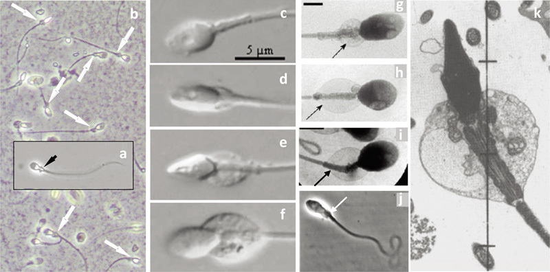
Montage of micrographs of (a) a human testicular spermatozoon (fixed in spermatocoele fluid), (b) human ejaculated spermatozoa (fixed in semen), both in phase contrast optics, showing the presence of cytoplasmic droplets (arrows) (‘midpiece vesicles') at the neck and midpiece region (Cooper TG, unpubl. data). (c–f) Living ejaculated human spermatozoa in semen showing droplets of different size (differential contrast microscopy from Fetic et al.90 with permission), (g–i) in medium (X-ray microscopy from Chantler and Abraham-Peskir91 with permission), in cervical mucus (j) (phase contrast microscopy from Abraham-Peskir et al.93 with permission) and a section of a human spermatozoon fixed in seminal plasma (from Smith et al.97 with permission). In (k), note the expanded vesicles that may be part of the Golgi membranes.56 Droplets are located only on the midpiece and may extend along its entire length.
