
Figure 4.
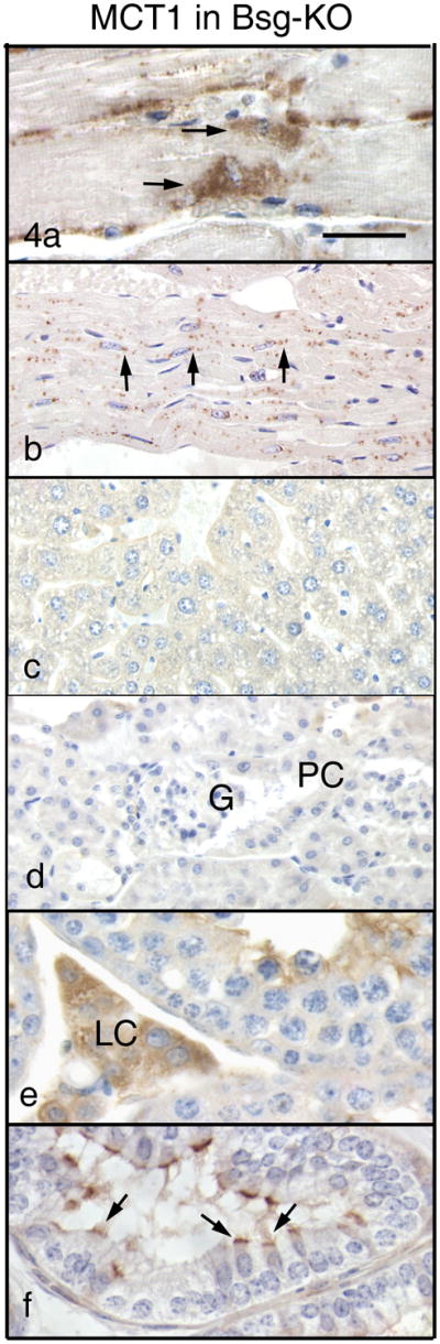
Immunohistochemistry of MCT1 in Group 1 of the Bsg-KO mice. Bar = 50 μm. In the skeletal (a) and cardiac (b) muscles, MCT1 disappears from the cell surface and cytoplasm but accumulates in the perinuclear area (arrows). No MCT1 immunostaining is seen in the liver (c) and kidney (d). G: gromerulus, PC: Proximal convoluted tubule. In the testis, Leydig cells (e, LC) remain positive for MCT1 in the absence of Bsg. In the efferent ductules (f), MCT1 disappears from the basolateral surface and accumulates on the apical border of ciliated cells (arrows).