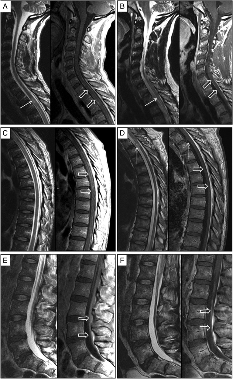
Figure 1.
Magnetic resonance imaging findings at presentation and after treatment. Sagittal T2-weighted and sagittal enhanced T1-weighted images of the cervical, thoracic, and lumbosacral spine upon presentation (A, C, and E respectively) and after treatment (B, D, and F respectively). Thickening and enhancement of the meningeal covers (hollow arrows in A and C) is noted upon presentation. This improves significantly on the follow-up examination (hollow arrows in B and D). Parenchymal cord involvement with abnormal T2-hyperintense lesion at T3 level noted upon presentation also improves significantly on follow-up (solid arrows in A and B). Enhancement of the cauda equina nerve roots (hollow arrows in E) is later replaced by nerve root clumping and enhancement (hollow arrows in F) suggesting the development of chronic arachnoiditis. Note the development of adhesions along the ventral aspect of the cord at T3–T4 level (white solid arrows in D).

