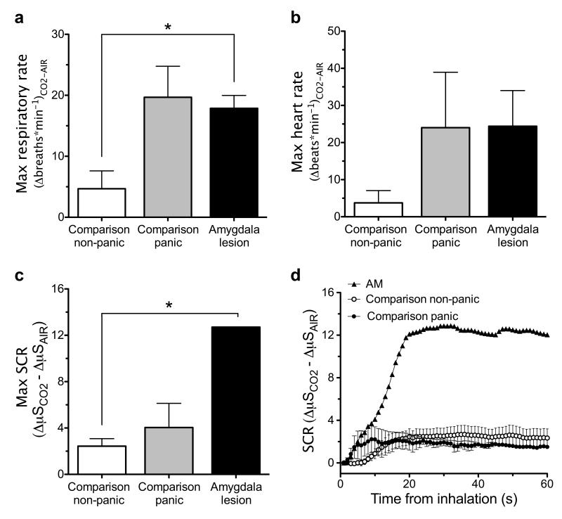
Figure 2.
CO2–evoked physiological changes. (a) Change from baseline in maximum respiratory rate during the first CO2 trial relative to the first air trial. Both the amygdala-lesion patients (n=3) and the comparison participants who panicked (n=3) demonstrated significantly higher increases in respiratory rate relative to the comparison participants who did not panic (n=9) (*p<0.05; Mann-Whitney U-tests). There was no significant difference between the amygdala-lesion patients and the comparison panickers. (b) Change from baseline in maximum heart rate during CO2 relative to air trials. Both the amygdala-lesion patients (n=2) and the comparison participants who panicked (n=3) demonstrated higher increases in heart rate relative to the comparison participants who did not panic (n=9). (c) Change from baseline in maximum SCR during the first CO2 trial relative to the first air trial. Patient AM demonstrated a significantly higher maximum SCR than the comparison participants who did not panic (*p<0.001; modified t-test). (d) Change from baseline in SCR during the first CO2 trial relative to the first air trial graphed during the first minute post-inhalation. Error bars represent the standard error of the mean.

