Figure 1.

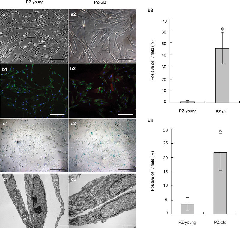
Determination of phenotype and ultrastructure of stromal cell cultures from normal prostate peripheral zone of young and old donors. (a1, a2) The PZ-old stromal cells were generally larger and more polygonal than the PZ-young cells. Scale bars=50 µm. (b1–b3) In double-label fluorescent immunocytostaining, all stromal cells were positive for prolyl-4-hydroxylase (green); a significant increase in the percentage (from 1.22%±0.97% to 45.67%±13.13%, *P<0.01) of α-SMA-positive cells (red) occurred with increased donor age. Nuclei are stained by DAPI (blue). Scale bars=50 µm. (c1–c2) SA-β-GAL staining showed more positive signals (blue) in PZ-old stromal cells; the quantitative data are shown in c3. *P<0.01. Scale bars=100 µm. (d1, d2) TEM ultrastructural analysis of stromal cell cultures derived from PZ-young and PZ-old donors. Compared to PZ-young stromal cells, PZ-old cells display increased dilated rough endoplasmic reticulum, Golgi complexes, prominent bundles of microfilaments and lysosomes in d2. Scale bars=2 µm. DAPI, 4′-6-diamidino-2-phenylindole; PZ, peripheral zone; α-SMA, alpha-smooth muscle actin.
