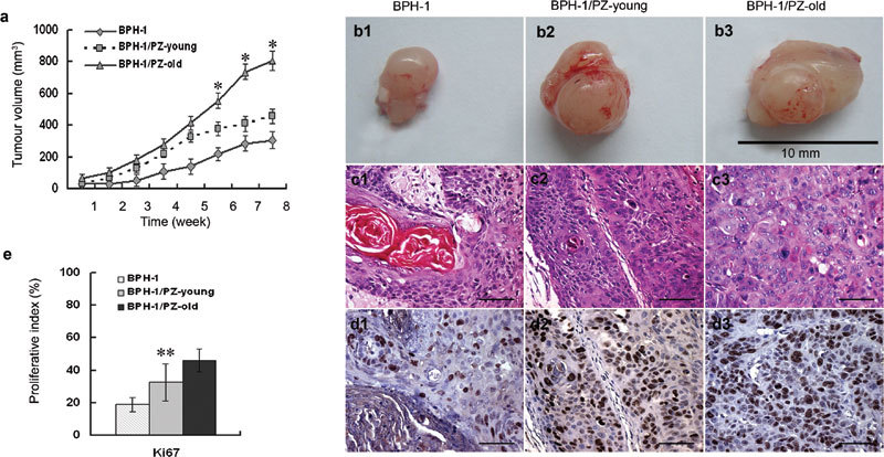
Figure 4.

Effect of prostate stromal cells on tumourigenicity of BPH-1 in athymic mice. (a) Growth curves of the xenografts. ‘Tumour' sizes were recorded weekly after inoculation until the experiments were terminated at 8 weeks. Tumour volume=width×length2×0.52. Data are presented as mean±s.d.. *P<0.01, compared with the other two groups. (b) Gross appearance of tissue xenografts prepared with BPH-1 cells alone or with mixtures of BPH-1/PZ-young and BPH-1/PZ-old cells, and harvested from male nude mice after 8 weeks of growth. (c) Histology of ‘tumour' sections from cocultured mouse models (H&E staining). Control BPH-1 cells inoculated alone showed the prominent appearance of many concentric ‘keratin pearls' (c1); ‘tumour' grafts from the BPH-1/PZ-young group showed small foci or cords of epithelium (c2), while cells in the BPH-1/PZ-old group displayed a much more undifferentiated phenotype with amphophilic cytoplasm and nuclear pleiomorphism with one or several prominent nucleoli (c3). Scale bars=40 µm. (d) Expression of Ki67 as assessed by immunostaining of ‘tumour' xenografts formed by BPH-1 cells (d1), BPH-1/PZ-young (d2) and BPH-1/PZ-old (d3) cocultured groups. Brown nuclear staining indicates a positive result. Scale bars=40 µm. (e) The expression ratio of Ki67 indicated that the proliferative status of cells in the BPH-1/PZ-old group was much higher than that of cells in the other two groups. **P<0.05, compared with other two groups. BPH, H&E, haematoxylin and eosin; PZ, peripheral zone.
