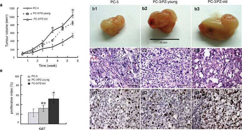
Figure 5.

Effect of prostate stromal cells on tumour progression of PC-3 cells in athymic mice. (a) Growth curves of the xenografts. Tumour sizes were recorded weekly after inoculation until the experiments were terminated at 6 weeks. Tumour volume=width×length2×0.52. Data are presented as mean±s.d. *P<0.01, compared with the other two groups. (b) Gross appearance of tissue xenografts prepared with PC-3 cells alone or with mixtures of PC-3/PZ-young or PC-3/PZ-old cells and harvested from male nude mice after 6 weeks of growth. (c) Histology of tumour sections from cocultured mice models assessed by H&E staining. All three groups (PC-3 cultured alone, PC-3/PZ-young and PC-3/PZ-old cocultured groups) displayed similar pleomorphic tumour cells. Scale bars=40 µm. (d) Tumour xenografts of PC-3 cells, PC-3/PZ-young and PC-3/PZ-old cocultured groups were stained with Ki67 antibody. Brown nuclear staining indicates a positive result. Scale bars=40 µm. (e) The expression ratio of Ki67 indicated that the proliferative status of cells in the PC-3/PZ-old group was much higher than that of cells in the other groups. *P<0.01, compared with other two groups. **P<0.05, compared with PC-3 group. H&E, haematoxylin and eosin; PZ, peripheral zone.
