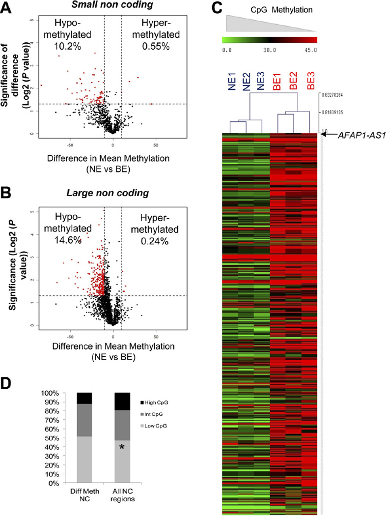
Figure 2.
Hypomethylation affects noncoding regions. (A and B) Volcano plots display differences in mean methylation between NE and BE samples (x-axis) and logs of P values between means (y-axis). Differentially methylated loci (red) reveal hypomethylation in both large and small noncoding regions. (C) Heat map displaying differential CpG methylation at all noncoding regions shows predominant hypomethylation. (D) Bar graph illustrating distribution of CpG densities according to differential methylation.

