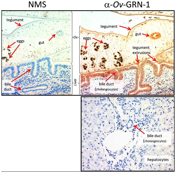
Figure 5. Immunolocalisation of Ov-GRN-1 in histological sections of adult O. viverrini in the bile ducts of experimentally infected hamsters.
The left panel was probed with IgG purified from control normal mouse serum (NMS); the right panel was probed with anti-Ov-GRN-1 IgG. The bottom panel shows a liver section from an uninfected hamster that was probed with anti-Ov-GRN-1 IgG. All three sections were stained with peroxidase staining revealed as a brown/rust colored deposit and Mayer’s Haematoxylin counterstained the nuclei in blue. Red arrows highlight the regions within the O. viverrini parasite and bile duct tissue that stained positive for Ov-GRN-1 75.

