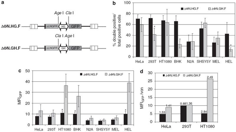
Figure 2.

Gene expression efficiency of the bidirectional promoter (a) Diagram of the foamy virus (FV) vectors with the two reporter genes (low-affinity nerve growth factor receptor (ΔLNGFR) and enhanced green fluorescent protein (EGFP)) cloned on either end of the promoter. GH denotes the alpha-galactosidase (GLA) to heterogeneous nuclear ribonucleoprotein H2 (HNRNPH2) orientation and HG the HNRNPH2 to GLA orientation of the promoter. A rabbit β-globin polyA was cloned in the 5′ end of ΔLNGFR. (b) Cell lines of diverse cellular background were transduced with the ΔΦN.HG.F or ΔΦN.GH.F FV vectors and the expression of EGFP and ΔLNGFR was analyzed by flow cytometry. The percentage of double-positive cells (coexpressing EGFP and ΔLNGFR) over the total number of reporter-positive (EGFP single positive+ΔLNGFR single positive+EGFP and ΔLNGFR double positive) cells from at least three individual experiments were plotted. (c) Comparison of promoter strength for each vector as determined by the total mean fluorescence intensity (MFI) of the EGFP protein. Values for both single- and double-positive cells were plotted. Mean values ±s.d. of at least three different experiments are shown. (d) Correlation of vector copy number determined by quantitative Taqman PCR and MFI in HeLa, 293T and HT1080 cells transduced with ΔΦN.HG.F or ΔΦN.GH.F FV viral vectors. Values above bars refer to average provirus copies per cell. (*P≤0.05).
