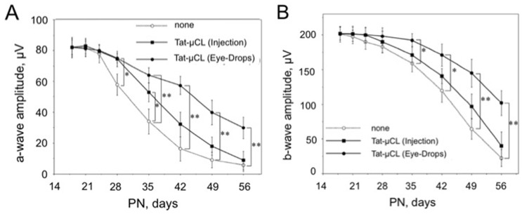
Figure 4. Effects of an intravitreal injection or eye drop applications of Tat-µCL on ERG in S334ter rats.
S334ter rats received an intravitreal injection of 2 µl of 20 mM Tat-µCL at PN 15 days (▪). Another group of S334ter rats received eye-drops containing 20 mM Tat-µCL from PN 13 to 55 days (•). Scotopic ERGs were recorded at PN 18, 21, 24, 28, 35, 42, 49, and 56 days. A) Mean amplitudes of photoreceptor-derived a-waves. B) Mean amplitudes of Müller cells-derived b-waves. Data are expressed as means ± standard deviation (n = 8 eyes (8 rats) per group). *P<0.05 and **P<0.01 versus the none-treated group (○) (t-test).

