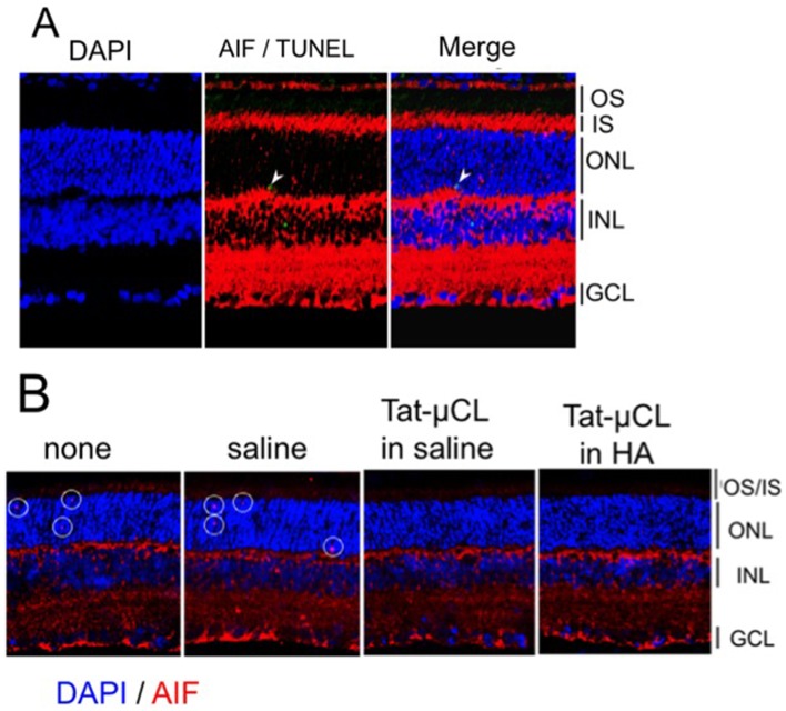
Figure 6. Determination of nuclear translocation of AIF in P23H rat retinas.
A) Eyes were enucleated at PN 40 days, and retinal sections were stained with AIF (red), TUNEL (green) and DAPI (blue). AIF was detected in photoreceptor cell nuclei. Arrows indicate localization of AIF in TUNEL-positive photoreceptor nuclei. B) Effects of eye-drop applications of Tat-µCL on nuclear translocation of AIF in P23H rats. Eye-drops containing saline (PBS), 1 mM Tat-µCL in saline, or 1 mM Tat-µCL in 0.1% HA were administered from PN 14 to 39 days. Eyes were enucleated at PN 40 days. Retinal sections were stained with AIF (red) and DAPI (blue). White circles indicate translocation of AIF inside photoreceptor nuclei (shown by pink color). Abbreviations: OS, photoreceptor outer segment; IS, photoreceptor inner segment; ONL, outer nuclear layer; INL, inner nuclear layer; GCL, ganglion cell layer.

