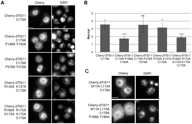
Figure 6. Identification of a cryptic PY-NLS-like motif in dTIS11 TZF.
(A) Plasmids coding for Cherry fused to WT or mutant versions of dTIS11 were expressed in S2 cells treated with LMB (10 ng/ml, 5h) and the localization of the fusions was observed by fluorescence microscopy. Bar = 5 µm. (B) The subcellular distribution of the fusions was quantified as described for Fig.2. Bars show the average of the nuc/cyt ratio ± s.d. *: p<0.05; ***: p<0.0001; ns: non-significant (U-tests). The significant differences between the nuc/cyt ratio of Cherry-dTIS11 C179A and those of the C179A/P189A/Y190A and R166A/R170A/K173A/K175A/C179A mutants were observed in two independent experiments. (C) Plasmids coding for Cherry fused to dTIS11 M111A L113A or dTIS11 M111A L113A C179A P189A Y190A were expressed in S2 cells and the localization of the fusions was observed by fluorescence microscopy. Bar = 5 µm.

