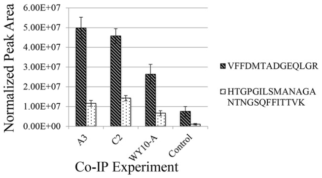
Figure 3. Normalized peak areas from two cyclophilin A peptides show enrichment in co-immunoprecipitation experiments using aphid proteins and purified CYDV-RPV.

Whole insects of genotypes A3, C2 and biotype WY-10A (efficient vector biotype recently collected from a field in Wyoming) were subjected to cryogenic cell lysis and protein extraction. The extracted proteins were co-immonoprecipitated with purified CYDV-RPV using anti-RPV antibodies. Two peptides from cyclophilin were enriched in gentoypes A3, C2 and the field collected biotype WY-10A as compared to the control co-immunoprecipitation with no virus (aphid proteins incubated with beads and antibodies). Peptide FFDMTADGEQLR (2+ precursor m/z 793.369) was higher in intensity than the HTGPGILSMANAGANTNGSQFFTTVK peptide (3+ precursor m/z 912.871). This is not a good indicator of differences in relative abundance between the two peptides, which could result from different ionization efficiencies. However comparison of peak areas for each peptide across the various samples is an accurate way to measure relative abundance of the peptide in each sample. Both peptides showed similar trends in the experimental co-IPs compared to the control. Both peptides were more abundant in the co-IP reactions with virus. Although the peak areas showed an overall lower abundance in biotype WY10-A, which might reflect a lower overall expression of cyclophilin in this biotype as compared to the lab-reared F2 genotypes, this difference was not significant using a Kruskal Wallis test.
