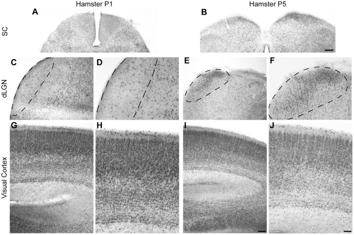
Figure 2. Expression of CB2R in the Superior Colliculus, dorsal Lateral Geniculate Nucleus, and Visual Cortex during Development in the Hamster.
Photomicrographs of coronal sections illustrating CB2R expression at P1 (A, C, D, G, H) and P5 (B, E, F, I, J) in the superior colliculus (SC) (A, B), the dorsal lateral geniculate nucleus (dLGN) (C–F), and the visual cortex (G–J). In panel C–F, dLGN has been outlined for better visualization. Scale bars: 200 µm (A, B); 100 µm (C, E, G, I); 50 µm (D, F, H, J).

