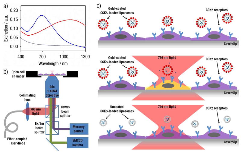
Figure 2.
(a) Extinction spectra of liposome preparations: uncoated liposomes (grey) and gold-coated liposomes with a plasmon resonance peak at 680 nm (blue) and at 1100 nm (red). Experimental samples were prepared and measured with equal quantities of lipids in solution and, therefore, presumably an equal number of liposomes per unit volume. (b) Schematic drawing of the inverted microscope setup for light-induced release and calcium monitoring. The 760 nm beam for light-induced release is produced by a pulsed fiber-coupled laser diode and is directed though a 60x objective to illuminate HEK293/CCK2R cells through an IR/VIS beam splitter. Indo-1 intensity from HEK293/CCK2R cells is monitored through the same 60x objective and imaged using an EMCCD camera. (c) Schematic drawing of light-induced release from gold-coated liposomes. HEK293/CCK2R cells are incubated with gold-coated liposomes, which only release and induce cellular activation when illuminated with 760 nm light. The microscope objective focuses the laser to obtain an activation area comparable to the surface area of the cell. Uncoated liposomes do not respond to the laser stimulus and do not induce cellular activation.

