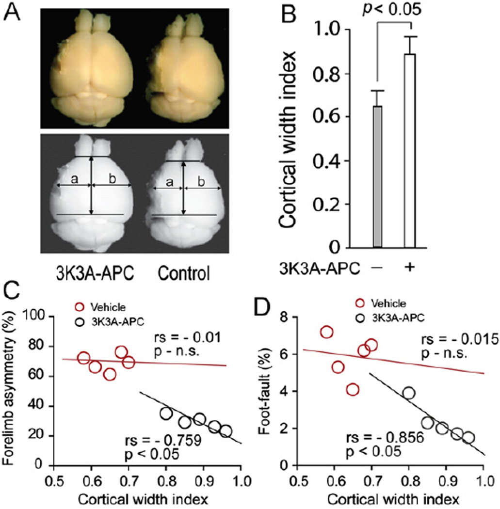
Fig. 4.
Effects of 3K3A-APC late multiple dose therapy on the cortical width index 14 days after permanent dMCAO. (A) Cortical cavitation (upper panels) and cortical width index measured using NIH Image J software (lower panels): a, maximum width from midpoint to edge of infarcted hemisphere; b, maximum edge from midpoint edge of noninfarcted hemisphere. F2r+/+ C57Bl6 mice treated with a multiple dose 3K3A-APC (0.8 mg/kg intraperitoneally at 12 h, 1, 3, 5 and 7 days after ischemia onset) or vehicle. (B) Cortical width index in mice treated with 3K3A-APC as above or vehicle. Regression analysis of forelimb use asymmetry test (C) and foot-fault test (D) versus the cortical width index at day 14. Mean±SEM, n = 5 mice/group. Pearson correlation coefficient (rs) and significance are indicated in panels C–D.

