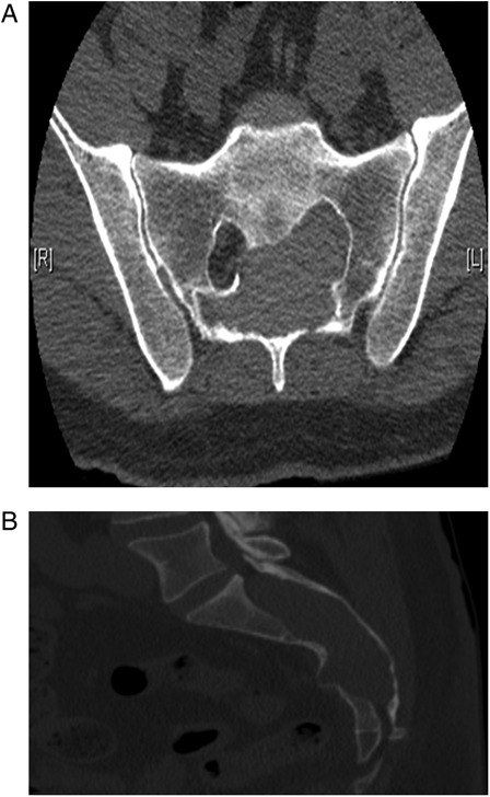
Figure 1.

(A and B) Axial and sagittal CT scan showed a large lobulated mass in the sacral spinal canal, osseous remodeling of the posterior vertebrae, and involvement of the left S1-3 foramina. A small portion of the mass extended through the right S2 foramen.
