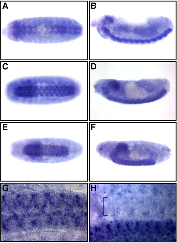
Fig. 3.

Sema-2b mRNA localization. A–F: Whole-mount photomicrographs of embryos processed for RNA in situ hybridization using Sema-2b probes. Anterior is to the left. A,C,E: Ventral views of an embryo centered on the VNC. B,D,F: Lateral views of the same embryos shown in the ventral view. A,B: Stage 13. C,D: Stage 15. E,F: Late stage 16. G: A dissected late stage-16 embryo focused on the VNC. H: A dissected late stage-16 embryo with both the VNC and the periphery represented. The bracket marks the ventral muscle domain.
