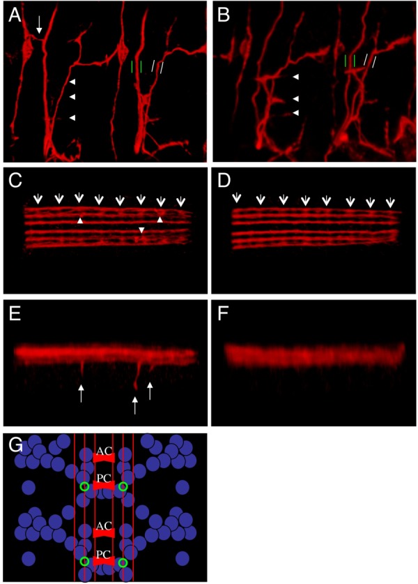
Fig. 4.

Sema-2b loss-of-function phenotypes. A–F: Confocal microscopy of late stage-16 embryos stained with mAb 1D4 antibody in red. A,C,F: f02042 homozygous embryos. B, D, E: f02042[Ex9] homozygous embryos. A,B: Views of two segments showing the ventral muscle domain and its innervation by ISNb. White arrowheads mark the normal innervation points by ISNb of the ventral muscles of one of the segments. Green parallel bars mark the ISN motoneurons and white parallel bars mark the SNa motoneurons to show their relative thicknesses in the two genotypes. White arrow shows an ectopic innervation from ISN to the LBD neuron. C,D: Ventral view of the VNC. White arrows mark the position of the posterior commissure of each segment and white arrowheads mark the position that ectopic ventral projections leave the medial fascicle. E,F: Lateral view of the same embryos as in C and D with arrows pointing to the ectopic ventral projections. G: A schematic of the mRNA expression of sema2b (blue), the mAb 1D4 fascicles (red), and the position where ectopic ventral projections are observed (green).
