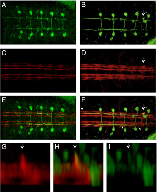
Fig. 5.

Visualization of Sema-2b-positive neurons in a Sema-2b loss-of-function background. Confocal microscopy of late stage-16 embryos expressing a sema2b-tau-myc transgene stained with anti-tau (green) and mAb 1D4 (red). Arrows point to an ectopic ventral projection. Arrowheads point to positions where the medial fascicle joins the lateral fascicle. Anterior is to the left. A, C, E: f02042[Ex9] homozygous embryos. B, D, F–I: f02042 homozygous embryo. A, B: Anti-tau. C,D: mAb 1D4. E,F: Merged z projection of the red and green channels. G–I: A lateral view of the VNC of a f02042 homozygous mutant embryo with an ectopic ventral projection.
