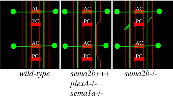
Fig. 6.

Schematic of Sema-2b phenotypes. A schematic of a stage-16 VNC with the three most prominent mAb 1D4 fascicles (red) and some of the Sema2b-Tau-Myc neurons (green) depicted. The wild-type VNC has Sema2b-Tau-Myc axons traversing in the middle mAb 1D4 fascicle after crossing through the anterior commissure. The middle schematic shows the effects of neuronal misexpression of Sema-2b and loss-of-function mutations in plexA and Sema-1a. The right schematic shows the effect of Sema-2b loss-of-function on the sema2b-positive neuron. The longitudinal axon is often seen moving from the middle to the lateral mAb 1D4-positive fascicle (shown on the right side of the embryo) and also to form an ectopic ventral projection, growing perpendicular to the plane of the longitudinal axons. anterior commissure (AC), posterior commissure (PC).
