
Figure 4.
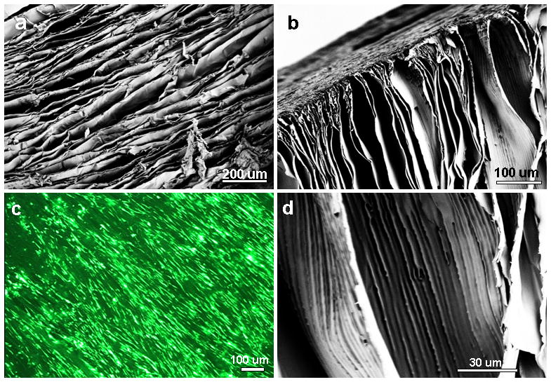
(a–b) Scanning electron microscopy images show aligned laminar channels within 3D silk scaffolds; (c) hMSC alignment and survival on silk scaffolds (Live-Dead staining); and (d) magnified image of thin scaffold wall with linear inner patterns.