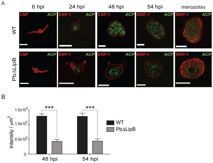
Fig. 5. PbΔLipB parasites have a less branched apicoplast in comparison to wild-type cells.
A. HepG2 cells were infected with WT or PbΔLipB sporozoites and the apicoplast was labeled using ACP-specific antibodies (labeling shown in green) at several time points throughout liver stage development. To visualize the developing parasite, primary antibodies (labeling shown in red) were used as follows: anti-CSP at 6 hpi; anti-Exp-1 at 30 & 48 hpi; anti-MSP-1 at 54 hpi and with mature merozoites (imaged at 62 hpi for WT parasites and 72 hpi for PbΔLipB parasites). White bar, 10 μm. B. Plot of intensity of apicoplast staining for WT vs. KO parasites sampled at 48 and 54 hi. Intensity was measured from the anti-ACP image and determined per μm2 of parasite after cropping the images for the parasite-specific areas (bounded by the parasitophorous vacuole). Results showed a statistically significant two-thirds reduction in apicoplast signal in KO compared to WT parasites (***p<0.001; Mann-Whitney U test, n = 12–18 per group). Additional images of parasites sampled at 48 and 54 hpi are provided in Figs. S4 and S5 respectively.

