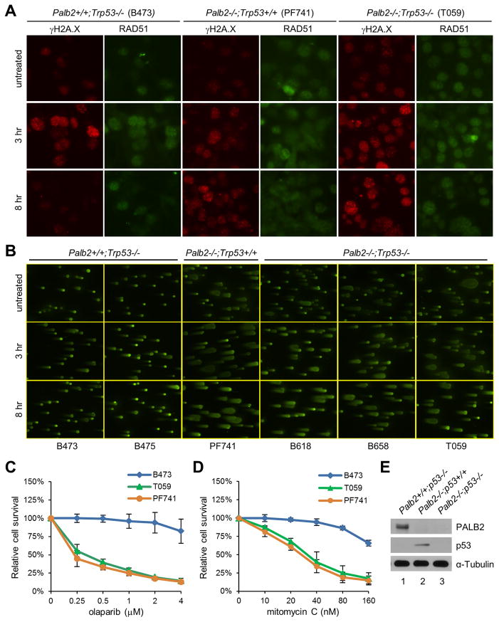
Figure 3.
DNA repair defect of Palb2-null tumor cells. A, γH2A.X and RAD51 foci formation before and after DNA damage induced by olaparib. Tumors cells were treated with 25 μM olaparib for 1 hr and the drug was then removed. Cells were fixed at 3 and 8 hr after drug removal and analyzed by IF. B, Levels of DNA breaks before and after olaparib treatment. Cells were treated as above, collected at the same time points and analyzed by neutral comet assay. C–D, Sensitivity of the tumor cells to olaparib and MMC. Cells were seeded in 96-well plates, treated with the drugs for 4 days and analyzed by CellTiterGlo assay. E, Western blots showing PALB2 and p53 protein levels in the tumor cells analyzed in C–D.

