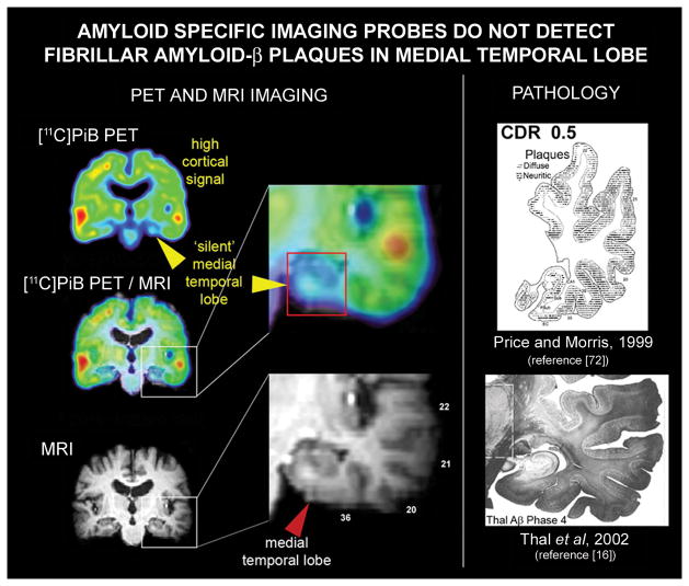
Fig. 4.
Mismatch between neuropathological results and ‘amyloid specific’ imaging probe signal in the medial temporal lobe. In AD medial temporal lobe structures have abundant amyloid-β deposit, both diffuse and neuritic plaques, comparable in density to those observed in the cortex as shown for temporal lobe by Price and Morris [72](upper image in the right column) and Thal and colleagues [16](lower image in the right column). Left column shows coronal view of the [11C]PiB PET image (upper image), of the MRI image from the same subject (lower image) and overlay of [11C]PiB PET image on MRI imaging for anatomical reference. Images in the central column show expanded temporal lobe structures on overlay of [11C]PiB PET image on MRI image (upper insert) and on the MRI image (lower image).The mismatch between amyloid PET imaging results and pathology in medial temporal lobe is demonstrated on an example of the AD [11C]PiB PET scan (upper image in the left row). The [11C]PiB PET signal is highly elevated throughout the cortex but is missing in the medial temporal lobe. Similar results have been reported for other “amyloid-specific” imaging probes. (This Figure contains parts of Figures from references [16] and [72]; with permission)

