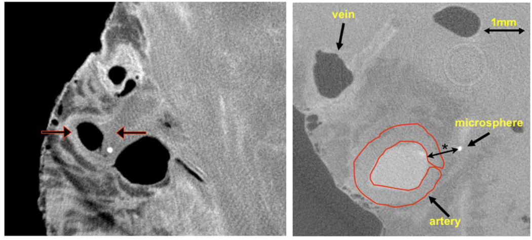
Fig. 1.
Left panel: Cryo-CT cross-section of epi-myocardial “biopsy” with epicardial artery, vein, and one microsphere in the arterial wall. The epicardium is the left interface to air and the fairly homogeneous region on the right is myocardium. The vein and arterial lumens contain air (dark in image) as these were allowed to drain at the time of harvesting. The left arrow indicates the opacified coronary arterial wall. The right arrow points to a region of the arterial wall without contrast in the wall due to the local vasa vasorum being occluded by an embolized microsphere (bright circle). Right panel: same CT image the coronary vessels with the region-of-interest (ROI) use to measure the CT value within the arterial wall

