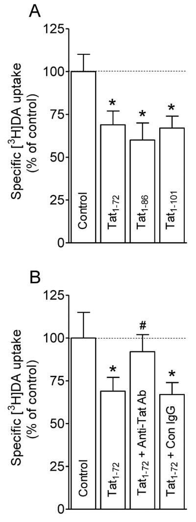
Figure 2.

Inhibition of DA uptake by released Tat from Tat-expressing cells. (A) CHO cells transfected with WT hDAT were preincubated in KRH buffer including 100 μl conditioned media collected at 72 h from cells transfected with plasmid Tat1-72, Tat1-86, Tat1−101 DNAs and vector alone (Control) followed by addition of [3H]DA uptake. * p < 0.05 different from control (Dunnett's Multiple comparison test). (B) Specificity of released Tat in inhibition of [3H]DA uptake. Conditioned media collected at 72 h from cells transfected with Tat1-72 were preincubated with anti-Tat antibody or isotype control IgG at 4°C for 3 h, followed by incubation with protein A/G – Agarose beads 4°C for 2 h. Media collected at same time from cells transfected with vector alone was used as control. Cells transfected with WT hDAT were preincubated in KRH buffer containing supernatants from the agarose-antibody-medium-beads complex, followed by [3H]DA uptake. Released Tat1-72 caused significant decrease in [3H]DA uptake, which was attenuated by immunodepletion with anti-Tat antibody but not isotype control antibody (one-way ANOVA followed by Tukey's multiple comparison test). * p < 0.05 different from control. # p < 0.05 different from Tat1-72 and Tat1-72 + Con IgG. (n = 4).
