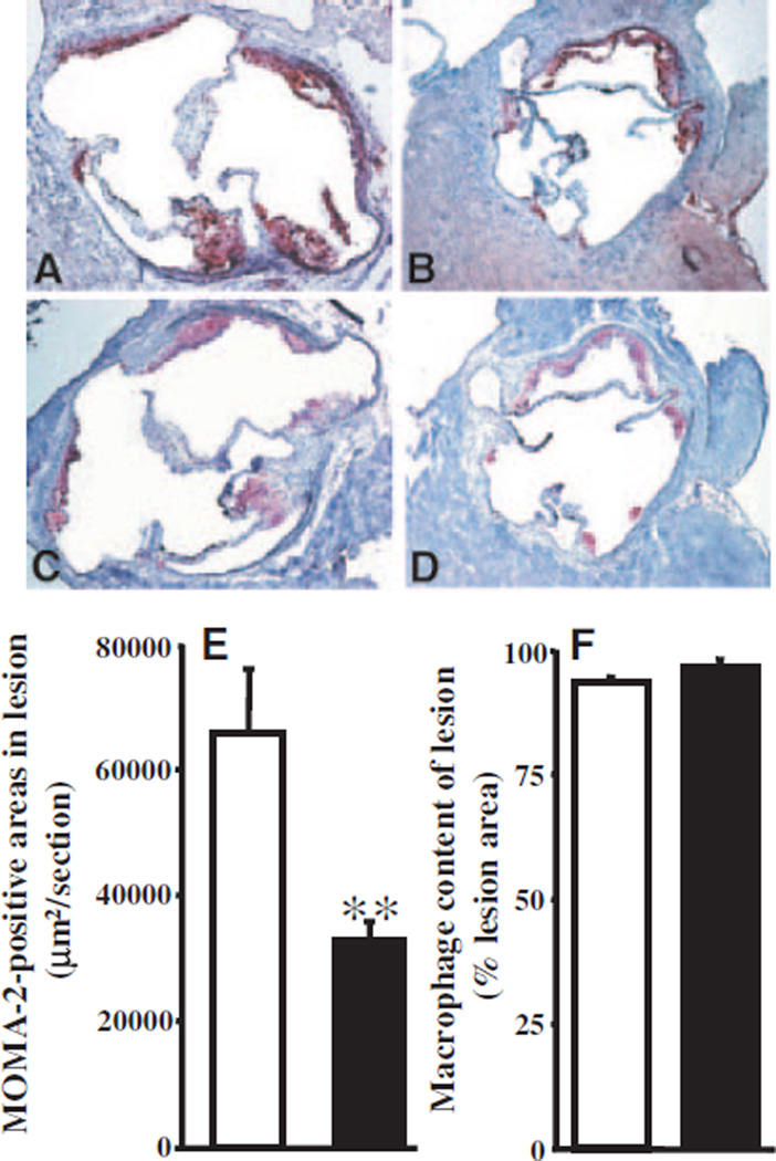
Figure 6.
Atherosclerotic lesions of male LDLRKO and PON3 Tg-1/LDLRKO mice fed the Western diet for 8 weeks. Representative sections of lesion areas at the aortic root region of male LDLRKO (A) and PON3 Tg-1/LDLRKO (B) stained with oil red O (red) and counterstained with hematoxylin (blue) are shown. Representative macrophage-positive areas of the atherosclerotic lesions at the aortic root region of male LDLRKO (C) and PON3 Tg-1/LDLRKO (D) mice stained with an antibody against MOMA-2 (red) and counterstained with hematoxylin are shown. Magnification is ×50 for A through D. MOMA-2 (macrophage)-positive areas (E) and macrophage content (F) of male LDLRKO (open bar) and PON3 Tg-1/ LDLRKO mice (filled bar) are shown. For each genotype, the mean and SE of macrophage-positive areas (E) or of macrophage content (F) of 20 sections from 5 mice are shown. **P<0.01 vs LDLRKO.

