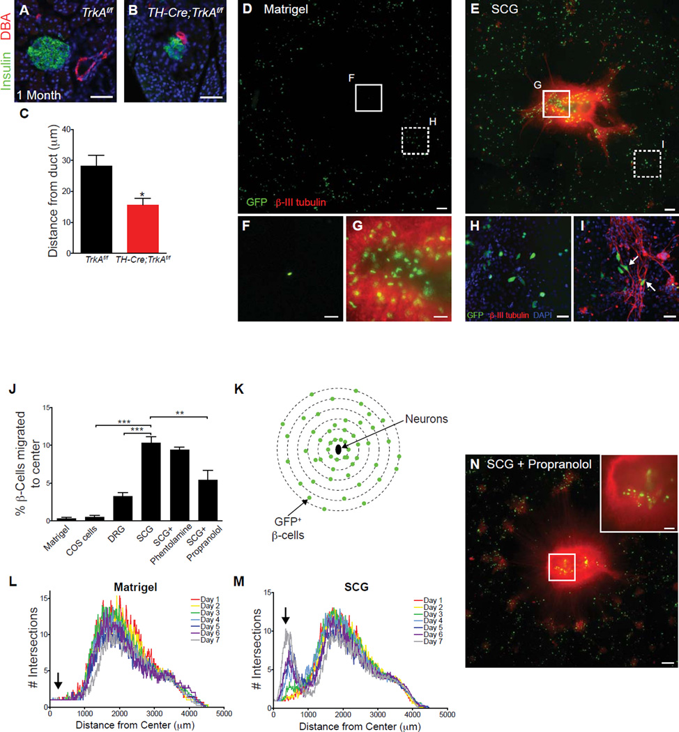
Figure 5. Sympathetic neurons promote β-cell migratory behavior through norepinephrine signaling.
(A–C) Ductal proximity of islets in one-month old TH-Cre;TrkAf/f mice. n=4 control and 3 mutant animals; mean ± SEM; *p < 0.05, t test. (D,E) Isolated β-cells exhibit directed migration towards sympathetic (SCG) tissue explants plated in the center of a Matrigel culture dish. Scale bars, 200µm. (F,G) Higher magnification images of the insets shown in D,E. Scale bars, 50 µm. (H,I) β-cells align along sympathetic axons and adopt polarized morphologies when cultured with neurons (arrows, I). Scale bars, 50 µm. (J) Percentage of β-cells that have migrated toward the center of Matrigel cultures containing COS cell aggregates, DRG or SCG explants with or without adrenergic antagonists after 7 days. n= at least 3 independent experiments; mean ± SEM; **p < 0.01, ***p < 0.001 by one-way ANOVA and Tukey’s post-hoc test. (K–M) Sholl analysis to quantify the distribution of GFP+ β-cells over 7 days in culture. The distribution of β-cells cultured alone does not change significantly over 7 days (L), while there is a gradual shift in the distribution of β-cells towards the center of the culture containing SCGs (M, arrow). Averaged profiles of four independent experiments are plotted. (N) β-cells exhibit decreased migration towards sympathetic explants in the presence of the β-adrenergic antagonist, propranolol (see J for quantification).

