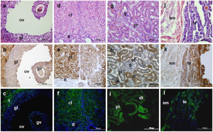
Fig. 3.
TSPO immunohistochemical staining and Tspo-AcGFP synthesis in the female urogenital system. The ovary (A–F; t, theca cells; tl, theca lutein cell; gl, granulosa lutein cells; gv, germinal vesicle; ov, ovarian follicles; cl, corpus luteum), kidney (G, H, I; g, glomerulus; pt, proximal tubules, dt, distal tubules; pl, parietal layer of male's glomerulus), and bladder (J, K, L; sm, smooth muscle; te, transitional epithelium) were examined using H&E staining (A, D, G, J), TSPO immunostaining (B, E, H, K), and AcGFP presence detected by fluorescence microscopy (C, F, I, L)

