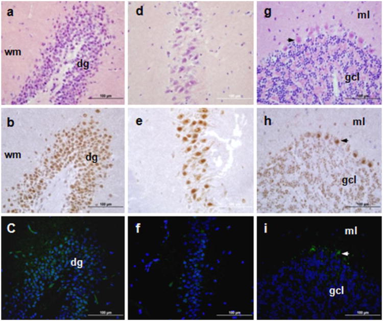
Fig. 4.
TSPO immunohistochemical staining and Tspo-AcGFP presence in the central nervous system. The hippocampus (A, B, C; dg, dentate gyrus, wm, white matter), cornu ammonis 3 (CA3) region (D, E,F), and cerebellum (G, H, I; arrows indicate Purkinje cells; ml, molecular layer; gcl, granule cell layer) were examined using H&E staining (A, D, G), TSPO immunostaining (B, E,H), and AcGFP presence detected by fluorescence microscopy (C, F, I)

