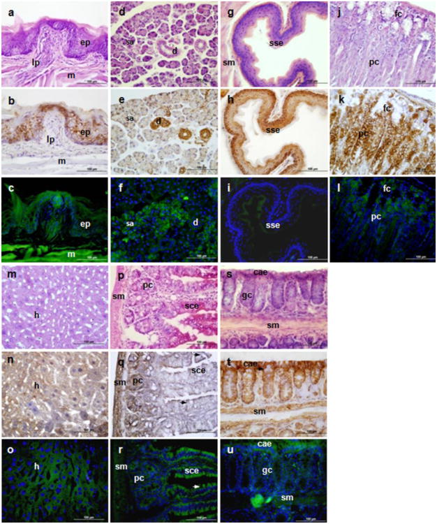
Fig. 7.
TSPO immunohistochemical staining and Tspo-AcGFP presence in the digestive system. The tongue (A, B, C; e, epethelium, lp, lamina propria, m, muscle), submandibular glands (D, E, F; serous gland, male mouse; d, duct, including intercalated portion and striated duct; sa, serous acini), esophagus (G, H, I; sse, stratified squamous epithelium), stomach (J, K, L; fc, foveolar cell; pc,parietal cells), liver (M, N, O; h, hepatocytes), small intestine (P, Q,R; sce, simple columnar epithelium, pc, crypt Paneth cell; sm, submucosa), and colon (S, T, U; cae, columnar absorptive epithelium, narrows indicate goblet cells; sm, submucosa) were examined using H&E staining (A, D, G, J, M, P, S), TSPO immunostaining (B, E, H, K, N, Q, T), and AcGFP presence detected by fluorescence microscopy (C, F, I, L, O, R, U)

