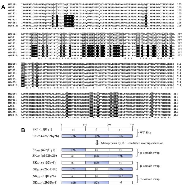
Fig. 1.
Structures of the proteins employed. (A). Sequence alignments of SK and SK2b from various GAS strains. Shown are amino-acid sequences of four SK1s (SKNS210, SKNS53, SKNS931, SKNZ131) and four SK2bs (SKAP53, SKNS223, SKNS455 and SKNS88.2) [21,32]. Strictly conserved residues in all SK1s and SK2bs are indicated with asterisks. The residues that are strictly conserved in one cluster, and different between clusters, are boxed (clear boxed for SK1, black boxed for SK2b). The sequence alignments were performed with ClustalX [33]. (B). Diagrams of the α, β, and γ domain-exchanged SK mutants from SK1 (NS931) and SK2b (NS88.2) used in this study. Domains were assigned according to the crystal structure of SKg [30].

