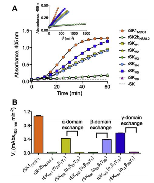
Fig. 4.
Activation of rGlu1-hPg by purified domain-exchanged SK mutants. (A). The rate of development of amidolytic activity monitored continuously by the absorbance at 405 nm at 37 °C using the chromogenic hPm substrate, S2251 (H-D-Val-Leu-Lys-pNA·2HCl). The control without rSK (dashed line) was performed under the same conditions. The inset shows the transformed plot of Absorbance at 405 nm vs. t2. (B). The initial velocities of hPm appearance for each rSK variant as calculated by linear regression from the linear regions of plots A405 nm vs. t2.

