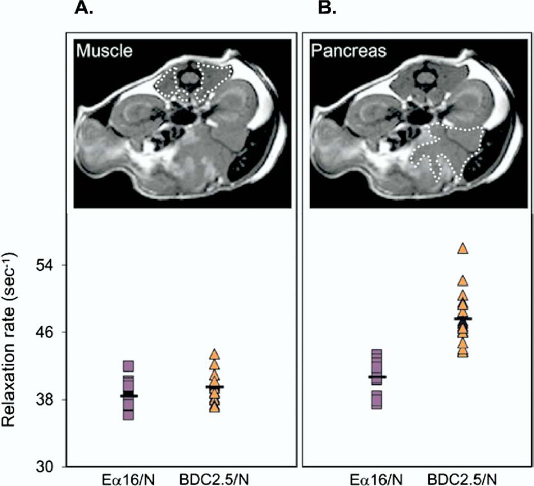
Figure 2.
Magnetofluorescent nanoparticles detected by MRI reveals microvascular alterations in a mouse model of insulitis. Twenty-four hours after injection of magnetofluorescent nanoparticles, MRI was performed in a BDC2.5/NOD mouse with insulitis and in an Eα16/NOD mouse without insulitis. A region of interest was defined on the MRI image over muscle tissue (A, upper) and over the pancreas (B, upper). The accumulation of magnetofluorescent nanoparticles was quantified in muscle tissue (A, lower) and in the pancreas (B, lower) in the 2 strains of mice. (Reproduced with permission from Moore et al,9 copyright ©2004 National Academy of Sciences, USA).

