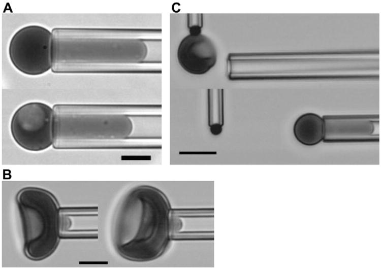
Figure 1.
Images of experimental procedures. (A) Measurement of area and volume. Cells are aspirated with sufficient pressure to form a spherical portion outside the pipette and a cylindrical projection within the pipette. The outer diameter of the spherical part and the length of the projection are used to calculate area and volume from the given equations. (B) Membrane shear modulus is determined by measuring the length of the cell projection in the pipette as a function of small increases in pressure. Cells with and without nuclei from an E15.5 sample are shown. (C) The stability of the bilayer skeletal interaction is measured by forming tethers from the cell surface. The cell is aspirated into the pipette and allowed to adhere to a target bead held at the tip of a microcantilever. The deflection of the cantilever from its rest position provides a measure of the tethering force. Changes in the aspiration pressure holding the cell in the pipette results in changes in the membrane tension. (A, B) Images obtained using a Nikon TE2000-E [436 nm monochromatic illumination, 100× 1.45 NA oil-immersion objective], Roper Quantem512SC digital camera, and Elements software (Nikon Instruments, Melville, NY); scale bar = 5 μm. (C) Images obtained with Olympus I×70 microscope [with 40 × 0.6 NA objective, 436 nm monochromatic illumination] and Hamamatsu C2400 CCD camera with background subtraction and brightness and contrast adjustments; scale bar =10 μm.)

