Figure 7.
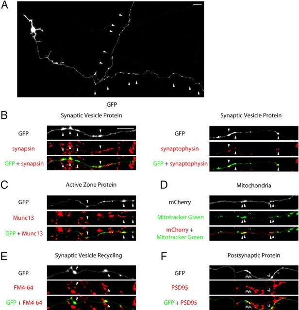
Characterization of maturing presynaptic boutons in primary neurons. A, Granule neurons were transfected with the GFP expression plasmid and subjected to immunocytochemistry using the GFP antibody at DIV12–13. A representative image is shown. Arrowheads indicate presynaptic varicosities along the granule neuron distal axon. B, C, Granule neurons transfected with the GFP plasmid were subjected to immunocytochemistry using the GFP antibody and the synapsin, synaptophysin, or Munc13 antibody. Arrowheads denote presynaptic varicosities colocalized with the endogenous synaptic vesicle protein synapsin or synaptophysin (B), or the active zone protein Munc13 (C). D, Granule neurons transfected with the mCherry expression plasmid were incubated with 100 nm of the Mitotracker Green dye for 30 min before imaging. Arrowheads denote varicosities colocalized with mitochondria as indicated by Mitotracker-positive puncta. E, Granule neurons transfected with the GFP expression plasmid were analyzed for synaptic vesicle recycling as in Figure 2E. Arrowheads denote coclusters of presynaptic varicosities with sites of FM4-64 dye uptake. F, Granule neurons transfected with the GFP plasmid were subjected to immunocytochemistry using the GFP antibody and the PSD95 antibody. Double arrowheads indicate presynaptic varicosities apposed to PSD95. Scale bars: 10 μm.

