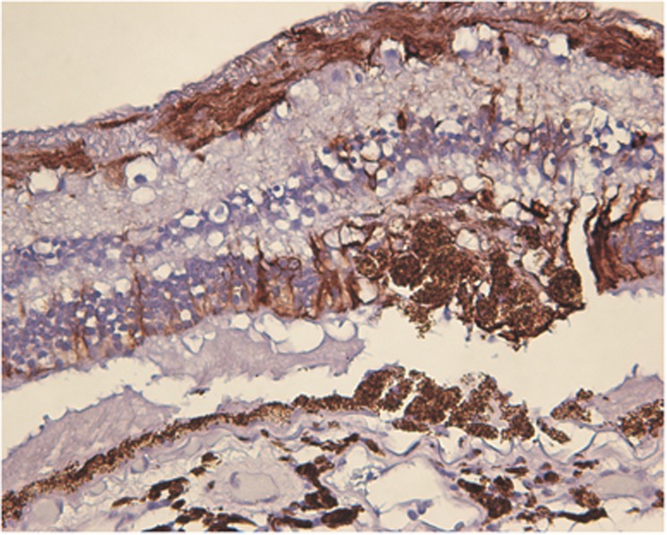
Figure 3.
Immunohistochemistry × 20 magnification with glial fibrillary acidic protein (GFAP), which stains intermediate filaments of astrocyte processes and activated Müller cells. There is subretinal fluid, suggesting these are likely the 1-month lesions. There are short vertical segments that stain positive for GFAP and extend from the INL to ONL, likely representing activated Müller cell processes or astrocyte gliosis.

