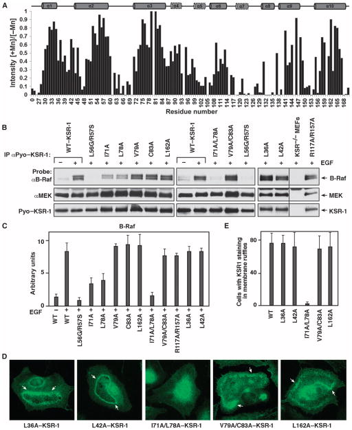
Fig. 6.
CC-SAM residues Ile71 and Leu78 mediate membrane binding. (A) Residues in α3 were tightly associated with micelles, as monitored by Mn2+ protection. Normalized 2D [1H,15N] HSQC peak intensities in the presence of MnCl2 (I[+Mn]) relative to those obtained in the absence of MnCl2 (I[−Mn]) are plotted for each residue. Peakswith ratios closest to 1 were most protected from the line-broadening effects of Mn2+. (B) ksr-1−/− MEFs stably expressing the indicated KSR-1 proteins were serum-starved and treated with EGF. Pyo–KSR-1 proteins were immunoprecipitated and examined for binding of endogenous B-Raf and MEK proteins. A representative blot from three separate experiments is shown. (C) Quantification of the B-Raf pull-downs is shown in (B). Error bars are the SD of the mean. (D) Serum-starved ksr-1−/− MEFs stably expressing the indicated KSR-1 proteins were stimulated with EGF, and the localization of the KSR-1 proteins was determined by immunofluorescence staining. Staining was performed in three independent experiments, and at least 200 cells were examined per experiment for each KSR-1 construct. Arrows indicate membrane ruffles. (E) Quantification of the localization results is shown in (D). Error bars are the SD of the average.

