
FIGURE 4.
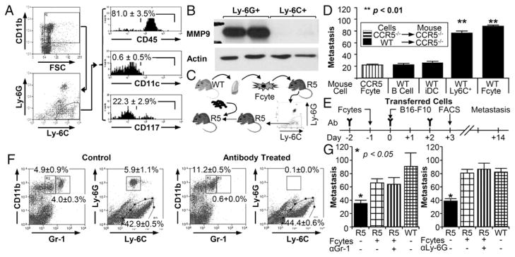
Ly-6C+, Ly-6Glo cells recruited by fibrocytes promoted metastasis. (A) Flow cytometric analysis of Ly-6Glo, Ly-6C+ cells. Gating strategy is on the left; histograms for CD45, CD11c, and CD117 are given on the right (n = 5). (B) Western blot analysis of Ly-6Glo, Ly-6C+ cells isolated from Ccr5−/− mice by flow sorting following injection with WT fibrocytes. (C) Experimental schema for defining function of Ly-6C+ monocytes. Ly-6C+ cells were isolated from Ccr5−/−(R5) mice 24 h after fibrocyte injection and transferred into R5 mice. Tumor cells were injected within 4 h of monocyte injection. (D) Bar graph shows metastasis in Ccr5−/− mice injected with Ccr5−/− fibrocytes or 5 × 10⁶ WT B cells, 1 × 10⁶ WT immature dendritic cells (iDCs), 2 × 10⁵ WT Ly-6C+ monocytes, or WT fibrocytes (n = 42). (E) Experimental schema for the depletion Gr-1 cells. Gr-1 or Ly-6G Ab was given on days −2,, 0, and 2 in relationship to the i.v. injection of B16 F10 cells (day zero). Fibrocytes (1 × 10⁵) were given on day −1; flow cytometry was performed on day 3. (F) Representative dot plots of pulmonary cells from mice treated with either Gr-1 or Ly-6G Ab. The graph on the left includes all cells (excluding debris by FSC/SSC); the graph on the right represents only CD11b+ cells. (G) Bar graph displaying the number of metastasis in mice treated with Gr-1 (n = 15) or Ly-6G (n = 31) Ab. Untreated Ccr5−/− mice (black) had statistically fewer metastases than all other groups. There were no significant differences in Ccr5−/− mice injected with WT fibrocytes.